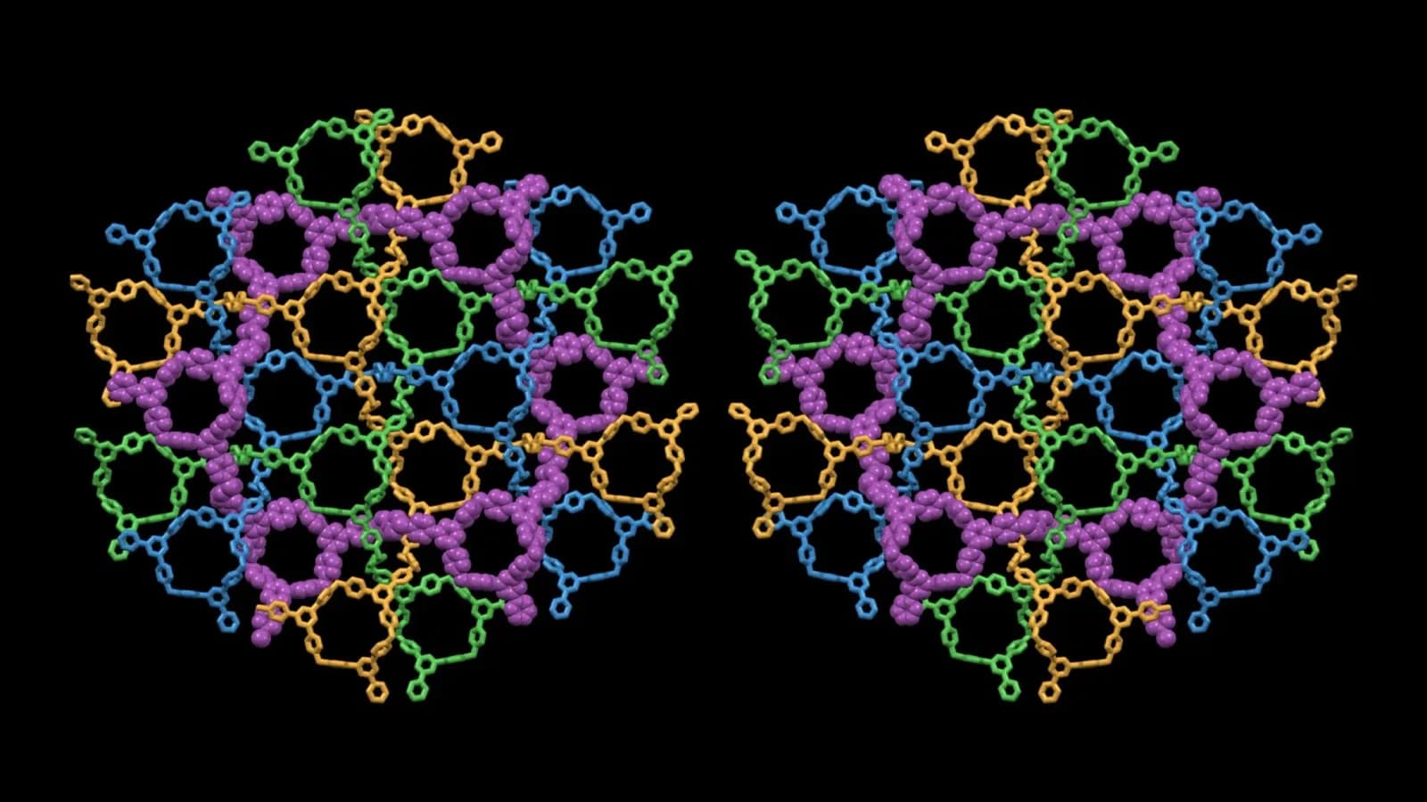
US team creates first 4D model showing how a microbe responds to changing light
Scientists have developed the first full four-dimensional model of a photosynthetic microbe to show how...

Scientists have developed the first full four-dimensional model of a photosynthetic microbe to show how a whole cell adapts to changes. The model follows the bacterium’s three-dimensional organization and the activity taking place inside it over time. It offers a detailed view of how cells coordinate internal processes as light levels change. The research, carried out by scientists at Pacific Northwest National Laboratory (PNNL) and the University of Illinois Urbana-Champaign, looks at Prochlorococcus marinus MED4, a small marine bacterium which uses sunlight to generate energy. This organism is a key player in ocean ecosystems and global carbon cycling.
“This study gives us a detailed view of how an entire photosynthetic cell responds,” said Margaret Cheung, computational scientist at PNNL and corresponding author.
Using cryo-electron tomography images, time-series data, physical models and high performance computing to bring together information about how the spatial organization of the cell relates to changes in gene, protein and metabolic processes. This allowed the team to see how different parts of the cell interacted.
A dynamic model of the cell
This model includes the fourth dimension of time to show how activity within the cell changes as the microbe transitions between light and dark. The team looked at cultures transitioning from dark to light to dark again, and also cultures in constant light or dark. This helped scientists understand how the cell responded to changes in conditions and how it was affected by the microbe’s biological clock.
“Coordinated measurements allowed us to test the model,” said Zan Luthey-Schulten, Professor of Chemistry at the University of Illinois Urbana-Champaign and corresponding author.
The research has shown that the spatial organization of proteins and other structures within the cell can affect the reliability of molecular interactions. The location of these structures help to reduce random variations in activity, meaning there is a strong link between the physical organization of the cell and regulation of its chemistry.
Switching carbon fixation on with light
For example, the CP12 protein regulates carbon fixation by binding to two enzymes involved in the process in the dark to form an inactive complex called the ‘dark complex.’ This means that in the dark the enzymes are unable to perform carbon fixation. When light is available, changes occur in the CP12 and enzymes causing the complex to disassemble to release the enzymes to begin carbon fixation again. This can happen quicker than the cell could make new proteins meaning the bacterium can rapidly respond to changes in conditions.
This study was published in the Biophysical Journal and was supported by the Department of Energy’s Biopreparedness Research Virtual Environment program. Some of the research was performed at the Environmental Molecular Sciences Laboratory (EMSL). Computing resources were provided by a 2024 Advanced Scientific Computing Research Leadership Computing Challenge award and the Tahoma high performance computing system at EMSL.
By looking at both the physical structure of the cell and activity over time, this model can help scientists understand how photosynthetic microbes respond to changes in conditions. This technique may provide scientists with a tool to understand how other cells regulate complex processes. Linking molecular activity to the whole living system. This may help guide future cell research.
Get the latest news delivered to your inbox.
Related articles